創薬による人と動物の健康長寿社会の実現 品種開発による持続可能な食糧生産 一本の染色体が次世代の医療や農業を支え、 人と地球の健やかな未来に貢献する。
このカテゴリーの記事はありません
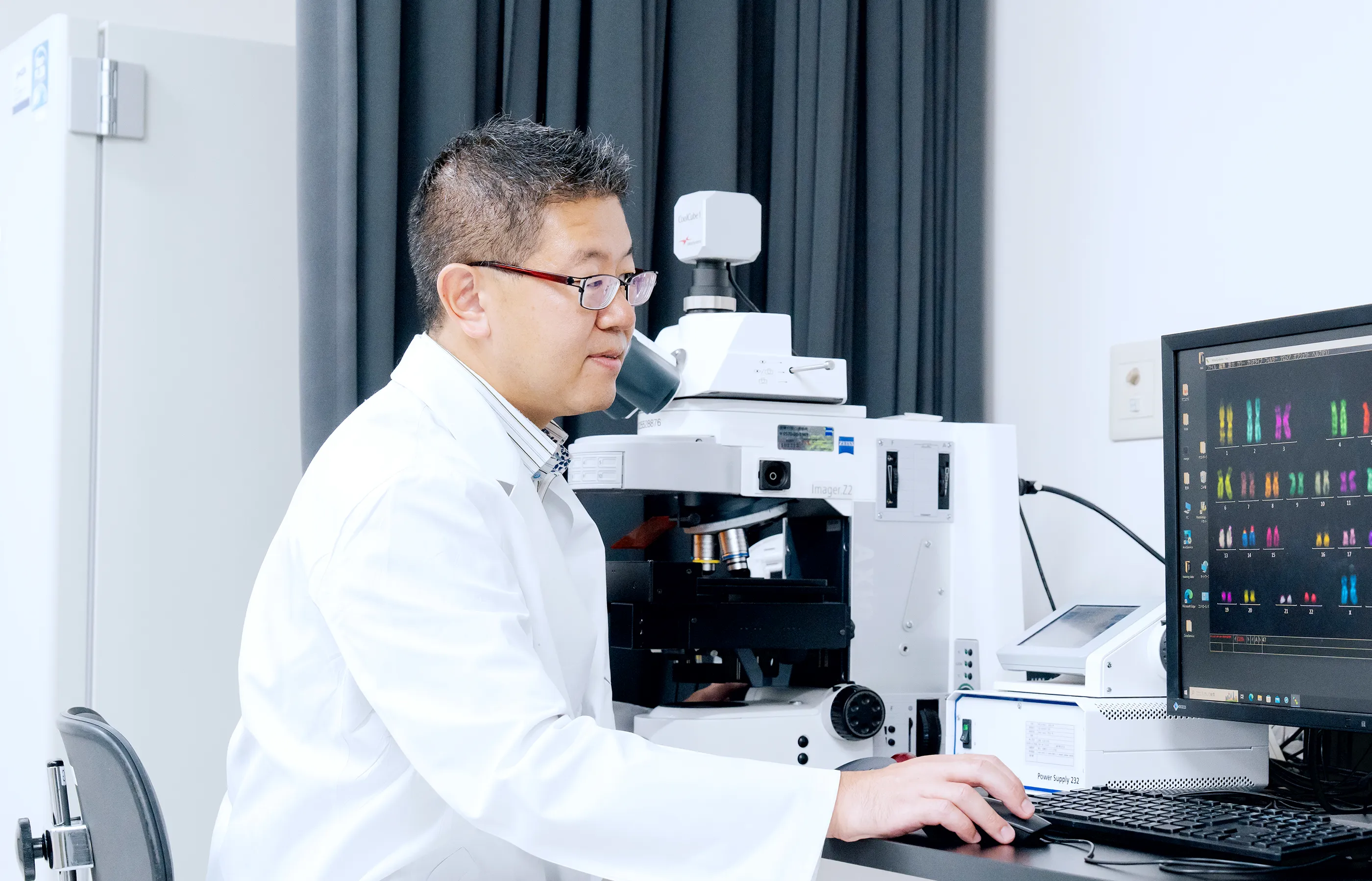
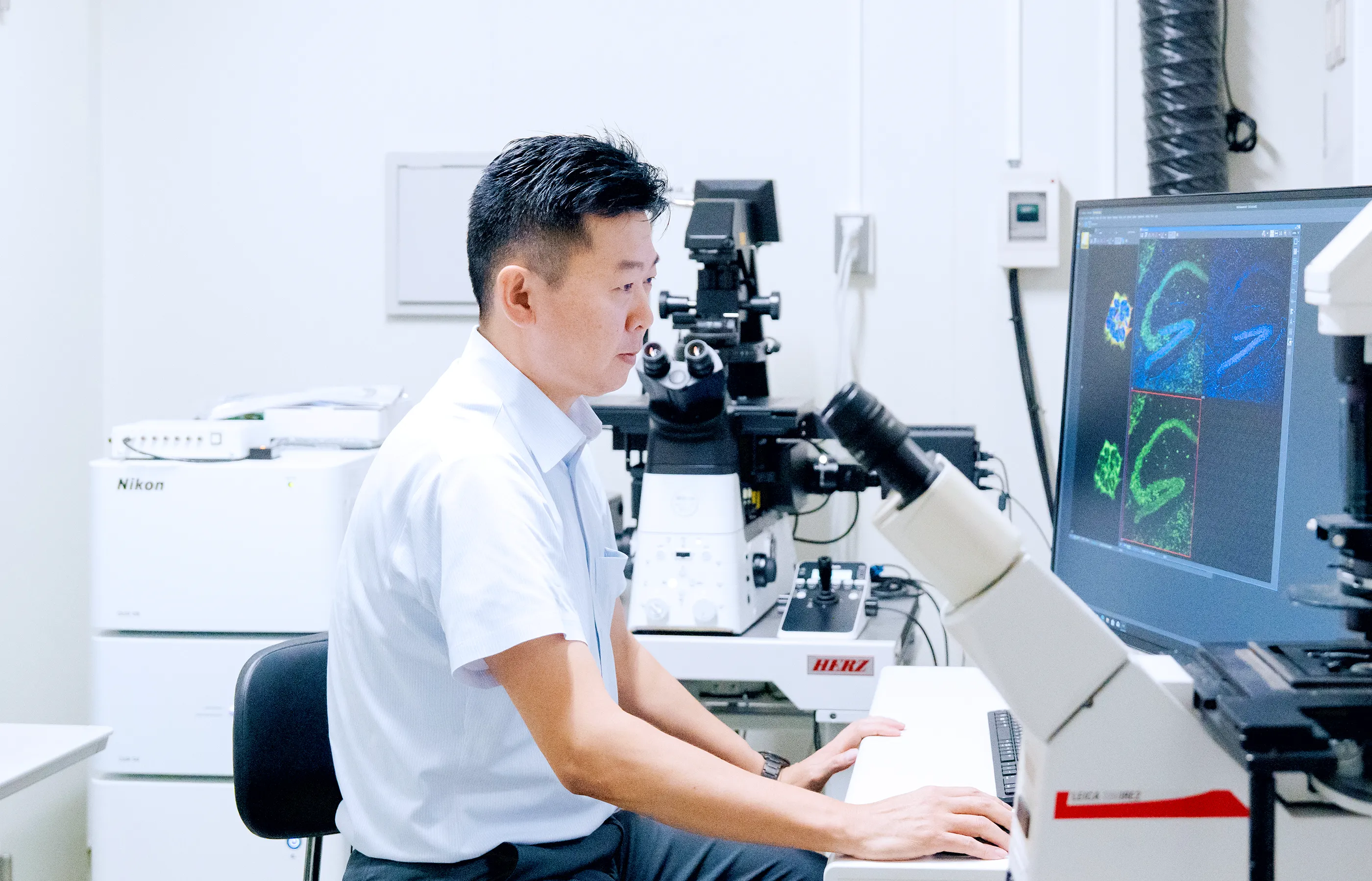
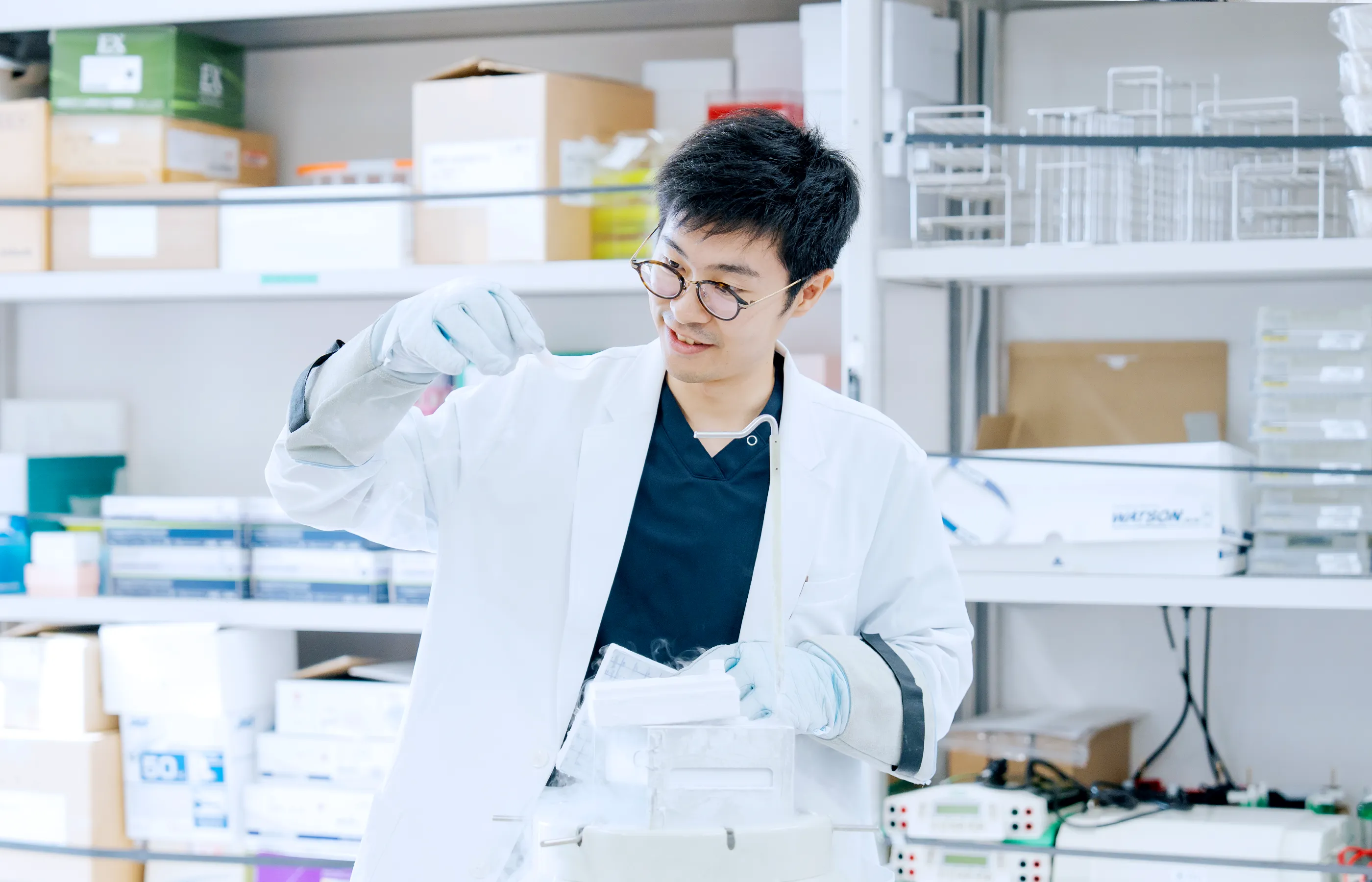
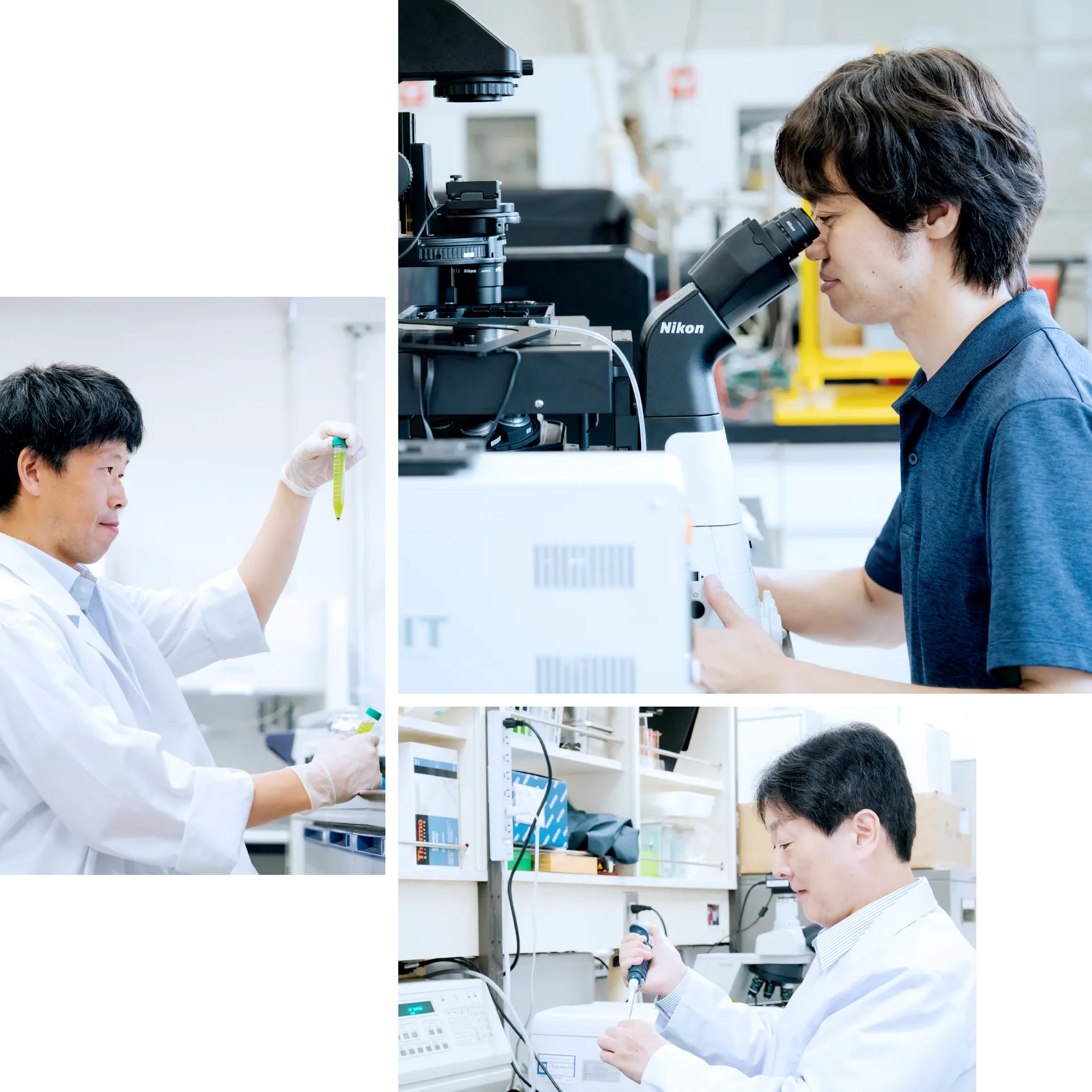
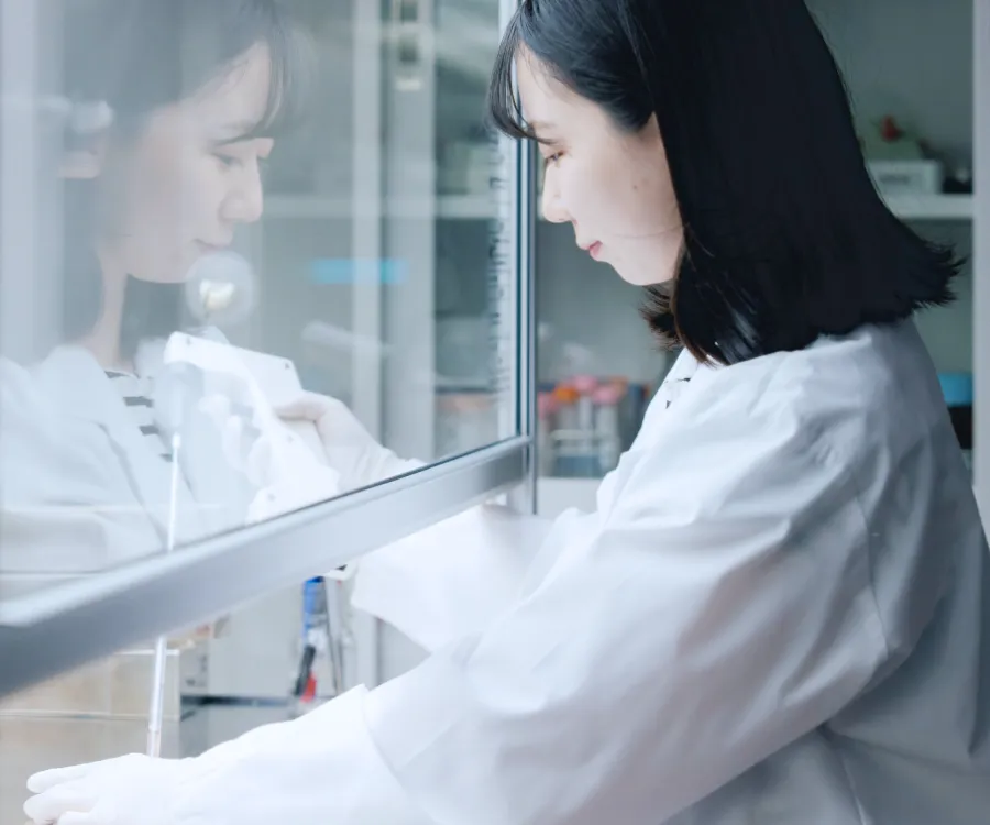
私たちの研究センターでは、最新の遺伝子解析装置や高性能顕微鏡を駆使して、染色体工学の革新的な研究を進めています。
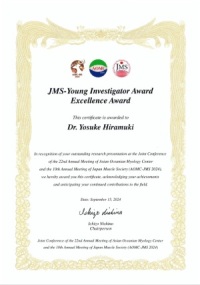
所属するすべての研究者の論文を紹介